Translate This Page
Eye Anatomy
The eye is an extraordinary and complex organ that allows us to 'see'. The brain actually does the 'seeing' - the eye is the 'central processor' that takes the information in the form of light waves and transmits the information into the brain.
The Orbit
The orbit helps to protect the human eye from injury and is comprised of seven bones:
- Ethmoid
- Frontal
- Lachrymal
- Maxillary
- Palatine
- Sphenoid
- Zygomatic
These bones converge and form a pyramid-shaped socket that points towards the back of the head. It is within this socket that the eyeball rests. Surrounding the eye within the socket is a layer of fat, cushioning the eyeball and allowing it to move smoothly within the orbit.
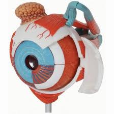
The Eye
(sometimes referred to as the eyeball)
The eyeball contains three layers:
- The outer layer, formed by the cornea and sclera
- The middle layer, holding the primary blood supply for the eye and containing the iris and pupil
- The inner layer, comprised of the retina
The eyeball also contains three chambers of fluid:
- Anterior chamber, between the cornea and iris
- Posterior chamber, between the iris and the lens
- Vitreous chamber, between the lens and the retina
The anterior and posterior chambers are filled with aqueous humour, which is a watery fluid that provides nourishment to the interior eye structures and helps to keep the eyeball inflated. The vitreous chamber is filled with a thicker fluid called vitreous humour, a transparent gel which is 99% water, which helps the eyes to stay inflated.
The Optic Nerve
As well as numerous blood vessels, the eye also contains the optic nerve. This runs from the back of the eyeball, through an opening in the orbit known as the optic foramen.
From here, the optic nerve connects to the brain and acts as a conduit, transmitting visual information into the brain. Other nerves within the eye carry non-visual information and send messages about pain or help to control motor activity within the eye.
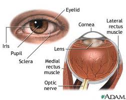
The Cornea
The cornea is the clear portion of the eye that covers the iris and the pupil and takes up about one-sixth of the eye. The rest of the eye (the scleral segment) is opaque. Several nerves and blood vessels run through the sclera, including the optic nerve. The cornea and scleral segment come together in an area called the limbus. This contains a great deal of blood vessels.
The Iris and Pupil
The iris and pupil are the most noticeable parts of the eye. The iris is the coloured ring of tissue that lies beneath the cornea and can be a range of colours, determined by genetics. The pupil is located in the centre of the iris and appears as a black hole that acts rather like a camera aperture, allowing light to enter the eye. This works in the same way as a camera, adjusting to control the flow of light into the eye. In bright conditions, the pupil closes down, reducing the amount of light entering the eye and protecting the delicate nerves from being damaged. In the dark, the reverse happens to allow what light there is to enter the eye.
Lens and Retina
Directly behind the iris is the lens. This focuses rays of light onto the retina, which is a light-sensitive nerve tissue that contains photosensitive cells called rods and cones. These convert light into electrical signals that are carried to the brain by the optic nerve.
Eyelids
Although often ignored, the eyelids play a crucial role in protecting the eyes. They help to protect the surface of the eye from scratches, dust and foreign objects and also help to lubricate the surface of the eye. When we blink, the eyelids carry secretions from the various glands across the eye.
The eyelid has several layers:
- A fibrous layer to provide stability
- A layer of muscle that controls the opening and closing of the eyelid. This layer can act incredibly quickly if the surface of the eye is vulnerable to attack, shutting the eyelid and protecting the surface of the eye using a reflex mechanism.
- A layer of skin that contains glands and the eyelashes, which act as filters to stop large foreign objects entering the eyes.
- The conjunctiva, which is a mucous membrane that connects the eyeball to the eyelid and the eyeball to the orbit.
Tear Glands
Round the eyelids. Tears are around 99% water and keep the cornea moist, as well as protect the delicate cells on the cornea. Each gland can have as many as 12 tear ducts. On blinking tears drain away via the puncta lachrymal, a small opening in the inner corner of the eyelid.
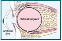
You can see some of the patients, who have undergone for procedure Custom Artificial Eyes, Silicone Eye Prosthesis, Ear Prosthesis, Facial Prosthesis, Magnetic Eye Prosthesis, Digital Eye, Treatment for mcirophthalmos & anophthalmos, with the Best ocularist in India, for the best Artificial eye and Facial Prosthesis, by Deepa D Raizada & Kuldeep Raizada, who are the world's Best Artificial eye maker and Facial Prosthesis Fabricator, at international Prosthetic Eye Center, India