


Enucleation is removal of the eye, leaving the eye muscles and remaining orbital contents intact. This type of ocular surgery is indicated for a number of different ocular tumors, in eyes that have suffered severe trauma, and in eyes that are blind and painful owing to other disease.[1]
Auto-enucleation (oedipism) and other forms of serious self inflicted eye injury are an extremely rare form of severe self-harm which usually results from serious mental illnesses such as schizophrenia.[2] The name comes from Oedipus, who gouged out his eyes in penance after having sex with his mother and killing his father.[3] Some patients have apparently been inspired by the Gospel of Matthew, which states: "...if the right eye offend thee, pluck it out and cast it from thee" (5:29).[4]
Classification
There are three types of eye removal
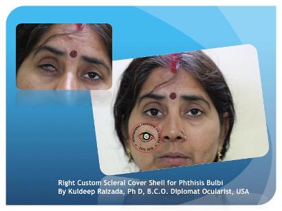
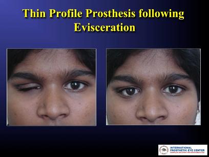
- Evisceration - removal of the internal eye contents, but the sclera is left behind with the extraocular muscles still attached.
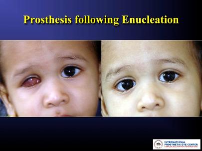
- Enucleation - removal of the eyeball, but the adjacent structures of the eye socket and eyelids remain. An intraocular tumor excision requires an enucleation, not an evisceration.
- Exenteration - removal of the contents of the eye socket (orbit) including the eyeball, fat, muscles and other adjacent structures of the eye. The eyelids may also be removed in cases of cutaneous cancers and unrelenting infection. Exenteration is sometimes done together with Maxillectomy which is removal of the maxilla or the upper jaw bone/cheekbone
Reasons for eye removal
- Cancer of the eye (retinoblastoma, melanomas, any other cancers of the eye or orbit)
- Severe injury of the eye when the eye cannot be saved or attempts to save the eye have failed
- End stage glaucoma
- Painful, blind eye
- In cases of sympathetic ophthalmia (inflammation of the eye) to prevent travel to other eye, in which, if untreated can cause blindness
- Congenital cystic eye
- In a deceased person, so the cornea can be used for a living person who needs a corneal transplant by a surgical operation called keratoplasty.
- Constant infection in a blind, or otherwise useless eye.
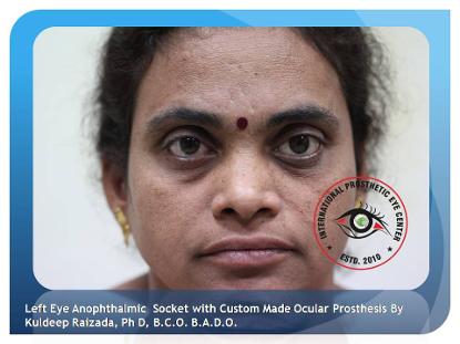
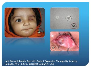
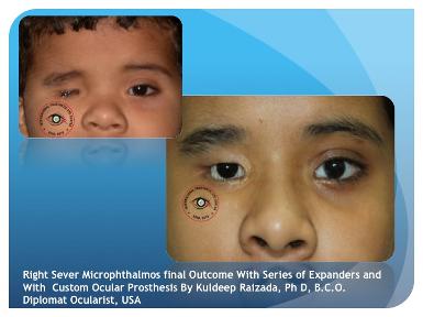
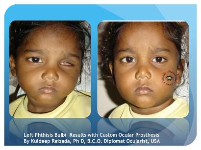
Orbital implants and ocular prostheses

Removal of the eye by enucleation or evisceration can relieve pain and minimize further risk to life and well-being of an individual with the above noted conditions. In addition, procedures to remove the eye should address the resultant appearance of the orbit. Orbital implants and ocular prostheses are used by the surgeon to restore a more natural appearance.
An orbital implant is placed after removal of the eye to restore volume to the eye socket and enhance movement or motility of an ocular prosthesis and eyelids. The eyeball is a slightly elongated sphere with a diameter of approximately 24 millimetres. To avoid a sunken appearance to the eye socket, an implant approximating this volume can be placed into the space of the removed eye, secured, and covered with Tenon's capsule and conjunctiva (the mucous membrane covering the natural sclera). Implants can be made of many materials with the most common being plastic,hydroxylapatite, metal alloy or glass.
Later, once the conjunctiva have healed and post-operative swelling has subsided, an ocular prosthesis can be placed to provide the appearance of a natural eye. The prosthesis is fabricated by an ocularist. Its form is that of a cupped disc so that it can fit comfortably in the pocket behind the eyelids overlying the conjunctiva that covers the orbital implant. The external portion of the ocular prosthesis is painted and finished to mimic a natural eye color, shape and luster. It can be removed and cleaned periodically by the individual or a care giver.
The two part system of orbital implant and ocular prosthesis provides a stable, and well tolerated aesthetic restoration of the eye socket. Although vision is not restored by removal of the eye with placement of an orbital implant and ocular prosthesis, a natural appearance can result. The implant can be moved by intact extraocular muscles that will track or move simultaneously with the other eye. The visible ocular prosthesis can couple with the orbital implant and thus move simultaneously with the other eye. The eyelids can move and blink over the prosthesis as well.